• facebook
  • twitter
  • linked in
  • google+
back top
Osteotomy
Knee Video Banner

Osteotomy

Osteotomy (literally ‘bone cutting’) is an operation in which a bone is cut to allow the bone to be repositioned; for example, to shorten, lengthen or realign. It may also be used to correct bones that have not healed properly following fractures or injuries. In the case of cartilage damage (whether recent or long-standing, such as in arthritis), it is also used to relieve pain and improve the environment for cartilage restoration in cases where poor alignment (‘malalignment’) is causing overload. This is analogous to a car that is out of alignment and has caused excessive tyre wear on one side.

The primary aim of osteotomy is to correct poor bone alignment and thus relieve pain, through the transfer of weight bearing from damaged to more healthy areas of the joint, such as in the knee. This hopefully increases the time to when a replacement joint is needed.

Due to the nature of the procedure, the operation is performed under general, spinal, epidural or regional anaesthetic.

Intended audience

This article is intended for anyone suffering from damage to their articular cartilage and their families who would like to find out about osteotomy, as well as anyone interested in cartilage problems.

What is Osteotomy?

As noted above, an osteotomy is an operation in which a portion of, for example, the tibia (shin) or femur (thigh) bone is cut and repositioned to restore proper weight bearing and bone structure. The primary aim of osteotomy is therefore to restore joint alignment by promoting more weight bearing on the side that still has healthy cartilage.

Surgeons often think of the knee as having three different areas:

  • The knee cap, as it rides on the end of the femur (patellofemoral compartment);
  • The inner knee between the femur and tibia (medial compartment); and
  • The outer knee between the tibial and femur (lateral compartments).

The most common use of osteotomy is to correct poor alignment (‘malalignment’) of the knee, which can occur at all of the above compartments.

In the two tibial and femoral compartments (medial or lateral), malalignment occurs if the limb is angled. This is often referred to as ‘bowleg’ or ‘knock-kneed’.

In the patellofemoral compartment, the patella rides along the outer aspect of the femur instead of the middle.

Figures

Tibial Tuberosity Osteotomy

How do you know if you are ‘malaligned’?

To find out exactly, your doctor needs to obtain specific X-rays. However, for the tibiofemoral compartments, some general sense can be obtained by standing in front of a long mirror and attempting to put your knees together:

  • If your feet touch but there is a space between your knees, you are bowlegged
  • If your knees touch but your feet are apart, you are knock-kneed

Both of these involve portions of the knee joint between the lower leg and/or the thigh (‘tibiofemoral joint’). The patellofemoral compartment is more difficult for you to ‘self evaluate’.

To precisely measure your alignment, your surgeon will obtain several X-rays. Tibiofemoral alignment is assessed with a long X-ray that shows the entire limb, from the hip joint to the knee and to the ankle.

In a normal hip to ankle (tibiofemoral) limb alignment, a line drawn on the X-ray connecting the hip and ankle joint is straight and runs through the middle of the knee. If the line is off-centre towards the inside (medial side) of the knee, this is known as ‘varus’ malalignment. If the line is off-centre towards the outside (lateral side) of the knee, this is known as ‘valgus’ malalignment. Any changes in alignment, whether varus or valgus, affect the distribution of the loads across the tibiofemoral compartment of the knee.

To assess the patellofemoral compartment, special images showing the patella riding on the femur are required, which can be more precisely measured with CT or MRI studies.

All of these malalignments alter the loading of the different compartments of the knee. These loads can be up to three times your body weight during normal walking, and up to six times your body weight during more strenuous activities. It is therefore not surprising that malalignment can be associated with both the development and progression of cartilage damage and ultimately osteoarthritis.

If the malalignment is corrected by bringing the line from the hip to the ankle back to an improved position at the knee, the pressure on the tibifemoral joint can be reduced. Similarly, if the patella is not precisely positioned in the femoral groove, centralizing the patella can reduce the patellofemoral joint stress. This may alleviate both pain and symptoms, and potentially improve the outcomes of cartilage repair procedures.

What are the different types of osteotomy?

Tibiofemoral osteotomy

Tibiofemoral osteotomy is typically recommended for malalignment. The extent is determined by the amount that the line is off-centre. Many surgeons consider that malalignment of more than 3º to 5º could benefit from realignment. Typical considerations involve one or more of the following circumstances:

However, the decision to perform surgery depends on your individual circumstances and discussions with your surgeon.

Osteotomy for bowed knees

If your surgeon has decided you have bowed knees (varus) and the problem is within the medial (inside) tibiofemoral compartment of the knee, your surgeon may recommend an osteotomy that corrects the knee alignment.

In most cases of varus knees, symptoms and problems are only on the inner aspects of your knee, thus the osteotomy will be performed on the tibia (shin bone), just below the knee. There are two ways to alter the alignment:

  • On the outer (lateral) tibia
  • On the inner (medial) tibia

As these cuts (osteotomy) are near the knee on the tibia, surgeons call this a ‘high tibial osteotomy’. In other words, it is high on the tibia relative to the ankle.

Lateral high tibial osteotomy

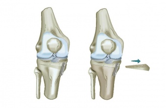
In this technique, the surgeon makes two angled cuts on the outer (lateral) tibial and remove a pie-shaped wedge of bone. The cut ends are then moved together (closed like a book) and fixed with metal (often a plate and screws). As this closes the pie wedge opening, surgeons call this a ‘closing wedge high tibial osteotomy’.

Medial high tibial osteotomy

In this technique, a single cut is made in the inner (medial) aspect of the knee and the cut edges are slowly pushed apart, like opening a book. Once the opening is sufficient to correct the alignment, the tibia is held in this new position, often with a plate and screws.

The opening might be filled with bone (from elsewhere in your body, from donor bone or a synthetic bone substitute). As this technique opens a wedge, surgeons call this an ‘opening wedge high tibial osteotomy’.

Osteotomy for knock knees

If your surgeon has decided you have knock knees (valgus) and the problem is within the lateral (outside) tibiofemoral compartment of the knee, your surgeon may recommend an osteotomy that corrects the knee alignment.

In most cases of valgus knees, with symptoms and problems only on the outer aspect of your knee, the osteotomy will be performed at the femur just above the knee. There are two ways to alter the alignment:

  • On the outer (lateral) femur
  • On the inner (medial) femur.

As these cuts (osteotomy) are near the knee on the femur, surgeons call this a ‘distal femoral osteotomy’. In other words, it is distal (‘away’), which in this case refers to the cut being further along the femur relative to the hip.

Lateral femoral osteotomy

In this technique, a single cut is made in the outer (aspect) aspect of the femur near the knee and the cut edges are slowly pushed apart like opening a book. Once the opening is sufficient to correct the alignment, the femur is held in this new position, often with a plate and screws.

The open area is often filled with bone (from elsewhere in your body, from donor bone or a synthetic bone substitute). As this opens a wedge, surgeons call this an ‘opening wedge osteotomy’ variant of the ‘distal femoral varus osteotomy’ (DFVO).

Medial distal femoral osteotomy

In this technique, the surgeon makes two angled cuts on the inner (medial ) femur and removes a pie-shaped wedge of bone. The cut ends are the moved together (closed like a book) and fixed with metal (often a plate and screws).

As this closes the pie wedge opening, surgeons call this a ‘closing wedge distal femoral osteotomy’. As it changes the knee from knock knee alignment or valgus, by moving it the other way (varus), surgeons also call this a DFVO, of the wedge-closing variety.

Osteotomy for malaligned knee caps

If your surgeon has decided you have malaligned kneecaps and the problem is within the patellofemoral compartment of the knee, your surgeon may recommend an osteotomy that improves the patellar alignment.

Patellar problems are unique and very complicated at times. There is often not “one” solution, but rather a patient specific approach involving more than one treatment. In addition to osteotomy, the patellofemoral problem may also require treatments such as balancing the soft tissues that connect the femur with the patella on the inner (medial) and outer (lateral aspects).

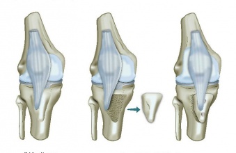
For the osteotomy portion of patellofemoral treatment, the cut (osteotomy) is made in the tibia in the region of the attachment of the patellar tendon (often a bony prominence on the tibia surgeons call the ‘tubercle’ or ‘tuberosity’). Putting these terms together, surgeons call this procedure a tibial tuberosity osteotomy (TTO).

The goal of tibial tuberosity surgery is to improve the patellar position within the femoral groove (much like placing a rope in a better position in a pulley) to decrease patellofemoral loading. The most commonly used TTOs with cartilage restoration/repair, move the tibial tuberosity either forwards (‘anteriorization’) or to move it forwards and towards the inside of the knee (‘anteromedialization’).

Anteromedialisation

Commonly termed AMZ or by the “inventor’s name, Fulkerson anteromedialization, the surgery begins with a sloped (angled) osteotomy (cut) of the tibial tuberosity. The patella tendon remains attached to this sloped bone cut. The tuberosity is then moved anterior and medial, bringing the patellar tendon attachment with it to a new position that both improves patellar tracking and decreases the patellofemoral forces.

Straight anteromedialisation

Some patients have patella and/or femoral cartilage problems, yet their alignment is normal. In these instances, the tuberosity may be moved straight forward. Historically, this required bone grafting, but newer techniques allow the osteotomy without bone graft.

What is my recovery like after osteotomy?

Obviously, this is a major surgery since the bone is cut. Your doctor will decide if this is performed in an inpatient or outpatient setting. For your return home, you will be switched from the surgical anesthetic to oral pain medication. Your surgeon will discuss ways to help decrease the occurrence of blood clots and infection.

Once home, during the early days, the goals are to decrease swelling with elevation (knee above your heart) and icing.

Rehabilitation after surgery is very gentle and specific to the surgery. It will depend on a number of factors, including your general health. A shared goal is early activation of the quadriceps muscle and the muscles around your hip, pelvis and core (seems illogical, but these muscles are very important for proper knee cap function. Weight bearing status and range of motion is very specific to your surgeon and the surgery performed, so discuss this with your surgeon.

Osteotomy healing is specific to the surgery and site of the procedure. The healing progress is usually assessed at 6 weeks. Sedentary work is often possible at this point, but heavy labour will require full healing of the osteotomy, excellent strength and near-normal function and is more commonly dictated by your cartilage restoration component of the surgery.

What are the advantages and disadvantages of Osteotomy?

What are the advantages of Osteotomy?

There are no activity restrictions following full recovery after an isolated and healed osteotomy, but other surgeries performed during the procedure, such as meniscal transplant, cartilage repair/restoration will have an impact on the final restrictions. In some instances, patients may be able to return even to high-impact sports, if they wish to do so understanding the risks of injury to their restoration.

As the procedure may delay the need for joint replacement (no guarantees), it is particularly beneficial in younger patients who have active lifestyles.

What are the disadvantages of Osteotomy?

There are many patients for which osteotomy will be unsuitable, including those with:

  • Poor cartilage on the opposite side of the poorly aligned bone
  • Inflammatory arthritis
  • Severely limited motion
  • Obesity
  • Joint dislocation
  • Osteoporosis
  • Nicotine use

When compared to partial knee replacement, osteotomy requires a longer period of rehabilitation, and the pain relief is not as predictable.

Swelling and numbness may persist in the weeks following surgery. There is also a risk of damage to nerves and blood vessels, although this is rare.

As with any open surgical procedure, there is a small risk of blood clots, poor healing and infection. Your doctor will discuss all of these risks with you prior to surgery.

Frequently Asked Questions (FAQs)

Will I have one leg shorter than the other after osteotomy?

The operated leg will be shorter by up to around 1 cm for closing wedge osteotomies and up to 1 cm longer in opening wedge osteotomies, but this is not usually noticeable after a few months.

Will osteotomy affect the success of a future total knee replacement?

While there was some previous thinking that this might be the case, newer systematic reviews of patients undergoing knee replacements after osteotomy have shown no significant impact on outcomes.

Further reading
  • Orban H, Mares E, Dragusanu M, Stan G. Total knee arthroplasty following high tibial osteotomy – a radiological evaluation. Maedica (Buchar). 2011;6:23-27.
Osteotomy (literally ‘bone cutting’)…
ICRShttps://cartilage.org/content/uploads/icrs.jpg
Thank You for Your vote

How useful did you find the information on this page?

Rating

5 (5 votes)